Cone Beam/ 3D X-Ray in Springfield, MO
Cone Beam Computed Tomography (CBCT), also known as a 3D X-ray, is used in conjunction with regular dental or facial X-rays. This uses a special technology that produces high-quality, 3D images of the dental structure, the soft tissues that make up the gums, the nerve paths, and the bone underlying the teeth with precision.
The images produced are then used to evaluate gum diseases, the bone framework of the face, nasal cavity, and sinuses, allowing more accurate treatment.
Uses of Cone Beam/3D X-Ray
Cone-beam CTs are used to detect complex oral problems, some of which include:
- Surgical planning for impacted teeth, i.e., the teeth that have not emerged due to lack of room or have grown in the wrong direction
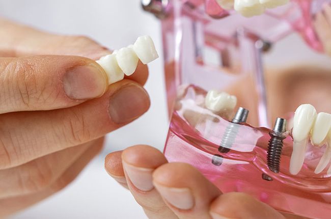
- Precisely placing dental implants
- Measuring and treating jaw tumors
- Determining teeth orientation and structure in an individual
- Helps in reconstructive surgery
- Determining whether or not a root canal is the most optimal choice for you
How Does a Cone Beam CT Scan Work?
The cone beam uses a rotating platform attached to an X-ray source and a detector. Due to the unique cone structure of the apparatus, the CBCT produces hundreds of planar projection images during a single rotation and allows a detailed field of vision. This is not possible in a traditional scanner. The fan-shaped beam stakes the individual image slices to create a complete 3D representation.
How Are You Prepared for a Cone Beam CT?
Cone-beam CTs are a very common, safe, quick, and easy imaging technique. Before the X-ray, everything that obstructs the field of view like metal objects, jewelry, eyeglasses, hair accessories, or hearing aids, should be removed. Removable dental appliances should be taken out before the procedure as well.
You will be asked to stand up tall, grab the hand rails and rest on a bite stick during the procedure. The C-arm of the apparatus rotates around your head and captures multiple images, which are then digitally combined to form the 3D image.
Advantages of a Cone Beam CT
- Cone beam CT has lower radiation exposure than conventional CT.
- It captures a wide variety of images from almost every angle that helps in an easier and more accurate assessment.
Please reach out to iTooth in Springfield, MO, to have a consultation with our dentists, Dr. Robbins or Dr. Fincel. Please call Dentist in Springfield MO at (417) 883-8515 or schedule an online consultation, and we'll guide you further.

